|
We are available around the clock.
|

|
|
| Our Medical Facilities |
- »Accident & Emergency
- »Anesthesia
- »Ayurveda/Homeopathy/Naturopathy
- »Bariatric and Metabolic Surgery
- »Blood Bank
- »Cancer Treatment
- »Cardiology
- »Cosmetic Surgery
- »Critical Care
- »Dental Surgery
- »Dermatology, Dentistry
- »ENT,Neurology,Neurosurgery
- »Gastroenterology and Hepatology
- »General,Chest Medicine,Endocrinology
- »General Surgery
- »Hematology
- »IVF and Endoscopy
- »Kidney Transplant
- »Laparoscopic Surgery
- »Medical Oncology
- »Neuro Trauma
- »Nephrology, Urology
- »Neurology
- »Neurosurgery
- »Nuclear Medicine
- »Obestetrics & Gynecology
- »Oncology
- »Opthalmology
- »Orthopedics and Joint Replacement
- »Pain Management & Palliative Care
- »Pathology
- »Pediatrics & Neonatology
- »Paediatrics Surgery
- »Physiotherapy
- »Psychiatry
- »Radiation Oncology
- »Radiology and Ultrasonography
- »Spine Surgery
- »Surgical Oncology
- »Urology & Stone care centre
- »Vascular Surgery
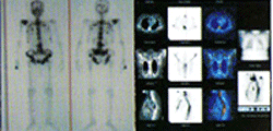
The Department of Nuclear Medicine, M. N. Budhrani Cancer Institute is equipped with a SPECT-CT scanner, The Infinia Hawkeye 4 Nuclear system from GE Healthcare which is the latest in hybrid scanners (i.e CT & SPECT Dual Head Gamma Camera).
It provides fully registered functional (SPECT) and anatomical information in a single image with a single examination. This helps us to determine the nature and precise location of the lesion. Fusion images from complementary modalities offer a more complete and accurate assessment of the disease.
Thyroid Scan
Indications:
- Diffuse Goitre / Multinodular Goitre
- Autonomous toxic adenoma
- Toxic Goitre
- Thyroiditis
- Solitary nodular Goitre
- Thyroid cyst
- Iodine-131 Whole Body Scan in cases of differentiated carcinoma thyroid
Parathyroid Scan
- Parathyroid adenoma/ Parathyroid hyperplasia
- Ectopic location of parathyroid adenoma
SPECT – CT enable localization of retro thyroid, intra thyroid and other
ectopic Parathyroid adenomas
Myocardial Perfusion Imaging / Stress Thallium
A normal stress MPI indicates a low risk (<1%) of developing any major event
in the next 1 year irrespective of the risk factors- Journal of Nuclear Cardiology
- Screening for IHD
- Triple vessel disease, prior to CABG.
- To look for ischemia in symptomatic post angioplasty patients
SPECT – CT provides for attenuation and scatter correction and hence accurate
interpretation of myocardial perfusion images.
Bone Scan

- Primary bone tumor
- Metastatic bone disease
- Fractures not detected by X-Ray
- Stress fracture v/s Shine splints
- Osteomyelitis v/s Cellulitis.
- Synovitis
- Avascular necrosis
- Metabolic bone diseases
- Pagets disease
- Fibrous dysplasia
Accurate localization by SPECT-CT helps differentiate benign from malignant
lesions.
Renal Imaging (DTPA & DMSA Scan)
- Bilateral / unilateral hydronephrosis
- PUJ obstruction / VUJ obstruction.
- Rental artery steno sis (Pre and post captopril studies)
- Congenital malformations of kidneys – DMMSA scan (Horse-shoe kidney, duplex kidney, ectopic kidney)
- Acute / chronic pyelonephritis –for cortical scars (DMSA scan)
- Vesicouretic reflux
- In renal transplant patients –ATN rejection, urine leak , lymphocoele formation, screening for donor
- Direct radionuclide cystogram for determining vesicoureteric reflux (VUR)
- GFR by “Plasma sampling methode” – a gold standard method for determining GFR (We are the first centre in pune doing this test)
HIDA Scan
- Acute calculus / acalculus cholecystitis
- Chronic cholecystitis
- Sphincter of oddi dysfunction
- Calculation of Gall Bladder ejection fraction
- Duodenogastric reflux
- Bile leak
- Biliary atresia v/s Neonatal hepatitis
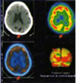
MIBI Brain SPECT
- To differentiate viable tumor tissue from radiation necrosis in postoperative, post radiotherapy patients of high grade gliomas

ECD Brain SPECT
- Detection of dementias
- Identifying epileptic focus
- Evalution of psychiatric disorders
- Assessment of stroke
Accurate functional information with anatomical localization by SPECT-CT
plats
a major role in further management
Other Scans
- Dacryscintrigraphy- painless, simple and patient friendly method to evaluate obstruction in the lacrimal system
- Salivary gland scintigraphy
- GI Bleed studies
- Meckle’s Scan (to determine functioning ectopic gastic mucosa)
- GE Reflux Studies
- MUGA Scan
- Lung ventilation perfusion studies:to detect pulmonary thromboembolism , for quantification studies prior to therapy
- Lymphoscientigraphy: To grade lymphoedema and decide upon the mode of treatment:medical/ surgical
Other Oncology Scans
- Scintimammography
- Tc-99m DMSA (V)for staging of Medullary Thyroid Carcinoma
- Tc-99m tektrotyd scan for somatostatin receptor imaging
- I-131 MIBG Scan
- Lu-177 DOTATATE Scans for neuroendocrine tumors
Infection Imaging
- Labelled WBC Scans
- Gallium 67 Scans
Therapeutic Nuclear Medicine
- Iodine -131 treatment in case if Graves disease/ Toxic goiters/ AFTN
- Iodin-131 treatment in post operative cases of differentiated with carcinoma thyroid
- P-32/Sm -153 / Sr-89 palliative pain therapy in cancer patient with multiple bone metastasis
- I-131 MIBG therapy in neuroblastomas
- Radiation Synovectomy in patients with Rheumatoid arthritis
- We are recognized by BARC for giving Radioactive Iodine Treatment for Ca Thyroid Patients
- Luthetium-177 DOTATATE therapy for well differentiated neuroendocrine tumours- We are the first facility in Maharashtra, approved by BARC for this procedure.
New Scans/Procedures
- Dopamine Transporter Imaging Tc99m TRODAT Scans.(To differentiate essential tremors from early parkinson’s).
- Somatostatin receptor imaging: Tc-99m HYNICTOC and In-111 Octreotide Scans for neuroendocrine tumours.
- Radiation Synovectomy for painful joints.
- Palliative pain therapy in bone metastasis.
- Lu-177 DOTATATE therapy in Neuroendocrine tumours.
- Iodine-131 MIBG Therapy in paraganglioma, metastatic pheochromocytomas, neuroblastomas.
For further information and appointment,
Please contact: 020-66099862
(Monday to Saturday,9 am to 5:30 pm)